Blogger: Bjørn Olav Haugen, Professor,
Centre for Innovative Ultrasound Solutions, Department of circulation and medical imaging

Angina is chest pain that occurs when the blood supply to the muscles of the heart is restricted. It usually happens because the coronary arteries supplying the heart become hardened and narrowed.
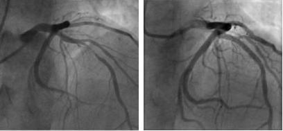
A coronary angiogram (CA) is an X-ray test done to find out if the coronary arteries are blocked or narrowed. If medication does not reduce the symptoms, it is usually recommended to do a coronary angiogram to help the cardiologist to see if you need treatment such as angioplasty with implantation of a stent (PCI), or coronary artery bypass surgery (CABG).
More than a million cardiac catheterizations with coronary angiography are performed annually in the US due to suspected coronary artery disease. According to data from The American College of Cardiology National Cardiovascular Data Registry, however, approximately 60% of planned coronary angiograms found no obstructive coronary heart disease.
Patients with heart attack (acute myocardial infarction) are usually sent to the catheterization lab, but in stable, non-acute patients with chest pain, unnecessary coronary angiograms should be avoided in order to reduce risk of complications in patients. This would also provide significant financial savings.
At the recent European Society of Cardiology Congress 2016 in Rome, two randomized trials were presented that could reduce the rates of coronary angiograms significantly.
The RCT CONSERVE Trial showed that using computed tomography (CT) to decide whether non-acute patients should undergo a coronary angiogram was both safe and cost-cutting, compared with a strategy of not using CT first to assess the need for a coronary angiogram. The new strategy led to an 86% reduction in coronary angiograms.
“The message from this trial is that, if we use coronary CT angiography as a gatekeeper to the catheterization lab in stable symptomatic patients with suspected coronary artery disease, we’ll reduce costs with sufficient safety,” says investigator Hyuk-Jae Chang, MD, PhD.
One important question raised by the expert panel during the session was why we should do CT angiography when the RCT PROMISE Trial showed that CT did not improve clinical outcomes compared with a so-called functional testing strategy (exercise electrocardiography, nuclear stress testing, or stress echocardiography).
The CE-MARC 2 Trial showed that a broader use of MRI or MSPECT scan, in low, intermediate and high risk patient groups, could reduce the rates of invasive coronary angiography (CA) that ultimately show no obstructive coronary disease.
Both studies underline the need for better strategies for assessing the pros and cons in terms of risk and usefulness of coronary angiograms in routine, non-acute, clinical practice.
Here at the Centre for Innovative Ultrasound Solutions (CIUS), we are working on the development of coronary anatomical and functional ultrasound imaging, safe techniques that we hope will provide a better tool for selecting patients that need coronary angiography and coronary angioplasty (PCI) or coronary artery bypass surgery (CABG).