Blogger: Eugene Kim, Post Doctor
MR Cancer Group, Department of circulation and medical imaging
This image won the prize for Best Scientific Image at the 8th International PhD Conference in Medical Imaging 2016:
Inspired by Vincent van Gogh’s signature bold, sweeping brush strokes, this contemporary piece evokes a meteor shower back-dropped by the majestic Milky Way stretching across the midnight sky.
It is a false-color image of a human breast cancer tissue section in which blood vessel walls and proliferating (growing/replicating) cells were labeled using immunohistochemistry. A digital image of the tissue section was acquired with a microscope. Then, a computer algorithm was used to segment the image into different classes or types of cellular and tissue components. Each color represents a different class (e.g., orange = blood vessel and yellow = proliferating cell).
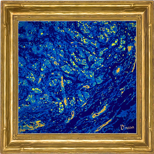
Technical details
The digital image of the human breast cancer tissue section was acquired at 40x with an Olympus VS120 slide scanner. K-means clustering was performed in MATLAB to segment the image into the different classes. This was part of a larger algorithm to automate the tedious task of counting proliferating blood vessels.