By: Ali Fatemi, PhD Candidate, Centre for Innovative Ultrasound Solutions (CIUS)
Despite constant improvements within the field of medical ultrasound, there are still a considerable number of difficult-to-image patient. Echocardiograms (heart images) taken from these patients do not have the quality that is needed for correct diagnosis. Therefore it is important to further improve the quality of echocardiograms.
Understanding the reasons behind low-quality images is essential to find methods to improve the quality. At the Centre for Innovative Ultrasound Solutions (CIUS), we have performed a systematic study to understand the mechanisms causing the problems in difficult-to-image patients.
In my previous blog post, we showed that the tip of a needle placed out of the imaging plane (the cross section of the object that is imaged), is seen in the ultrasound image if some part of the ultrasound signal hits the bones and gets deflected towards the needle. The question was then if there are some types of tissue in the body that can play the role of the needle and make noise by sending the deflected signal back to the ultrasound transducer.
We have now attempted to answer this question by performing different water tank experiments where we imaged an artificial heart chamber in the presence of two pieces of rib bones. In these experiments, we placed the bones at different angles relative to the ultrasound transducer and used different types of tissue such as skin, fat and lung to study the effect of these on the images.

Figure 1- Beamprofile measurement of an ultrasound transducer. a) without ribs, b) with ribs partly blocking the transducer, and c) with ribs at a different angle from b).
The results of these experiments show that depending on the angle between the ultrasound beam and the ribs, the ultrasound signal can be partly reflected either outside or inside the ribcage (see Figure 1). The measured energy from the transducer can be seen as a single beam in Figure 1a. In Figure 1b and 1c, the ultrasound signal is split in two parts after hitting the rib. Different angles between the transducer and the ribs in these two measurements results in reflection of signal outside the ribcage (above the bones) in 1b and inside the ribcage (below the bones) in 1c. Therefore, we can consider two cases:
Case 1: the reflection of signal is outside the patient’s chest.

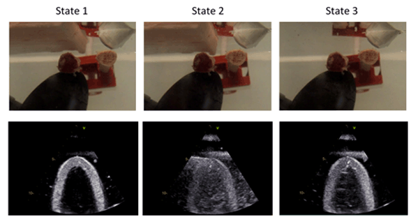
Figure 2- Watertank experiment with rib bones and a layer of fat. State 1) large distance between the ribs, state 2) reduced distance between the ribs, and state 3) the fat layer is removed.
This reflected signal can then hit the subcutaneous fat and travel back to the transducer. The received echoes from subcutaneous fat are then shown as noise over the image of the heart. We experimentally demonstrated this case by the setup shown in Figure 2. In this experiment, the bones were placed in a way that the reflection of the signal was outside the ribcage (same as in Figure 1b). The upper row shows three instances of this experiment, while the lower row shows the corresponding ultrasound images taken by the transducer. A layer of fat was placed over the ribs to simulate the effect of the subcutaneous fat inside the body. A clear ultrasound image of the heart chamber can be seen in State 1 where there is a big enough distance between the bones and therefore no reflection of signal towards the fat layer. In State 2, however, where the distance between the bones is reduced, the ultrasound image of the heart chamber is covered with a haze-like noise. Finally, in State 3, the fat layer is removed while keeping the distance between the ribs as short as in State 2. The haze-like noise is mostly removed from the ultrasound image in this state.
Case 2: the reflection of signal is inside the patient’s chest.

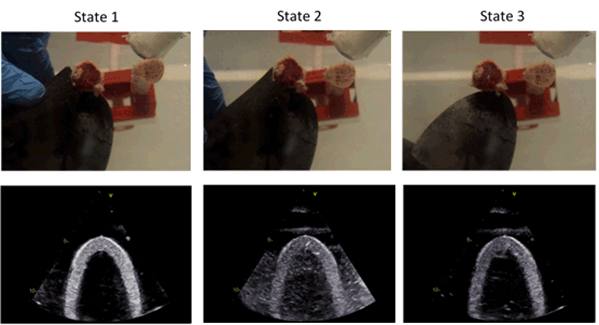
Figure 3- Watertank experiment with rib bones and a piece of sponge. State 1) large distance between the ribs, state 2) reduced distance between the ribs, and state 3) the sponge is removed.
In this case, the lung tissue can send the signal back to the transducer and cause the degradation of the image quality. This case was experimentally demonstrated by the setup shown in Figure 3. In this experiment, the bones were placed at a slightly different angle from the first experiment so that the reflection of the signal was inside the ribcage (same as in Figure 1c). The same steps were followed as in the first experiment, but instead of the fat layer over the ribs a piece of sponge was placed under the ribs and close to the heart chamber. The sponge simulates the effect of the lungs in the body because of the air trapped inside it. Similar ultrasound images as in previous experiment can be seen here.
The results of these experiments confirm that the deflection of ultrasound signal by the ribs in unwanted directions followed by a second reflection from either fat layers or lung tissue can generate noise in echocardiograms. This knowledge can be applied to implement new techniques in order to improve the quality of echocardiograms in difficult-to-image patients.
