Bloggers: May-Britt Tessem
and Morten Beck Rye
As we speak there are no accurate methods to diagnose potentially dangerous prostate cancer in an early stage of cancer.
From a pathologist’s point of view, aggressive cancers look totally similar to harmless subtypes in the beginning of development. As a consequence, the patients will be at high risk of overtreatment in the majority of cases where prostate cancer is detected. We urgently need new tools and markers to sort out the potentially dangerous types of prostate cancer from the non-dangerous in early disease. Most importantly, this will save the patients from reduced quality of life due to unnecessary surgical interventions, and also be economically beneficial for society.
With this background, the MR Cancer group (ISB) in cooperation with the Bioinformatics and gene regulation group (IKM) and Dept. of Urology at St Olav, investigated possible subgroups of prostate cancer based on expression of specific sets of genes. We were especially interested in gene sets which could assist clinicians in prognostic decisions making, and which could be used in addition to existing clinical parameters. To sort out dangerous prostate cancer in early disease progression, will aid in planning of a personalized treatment strategy for each patient.
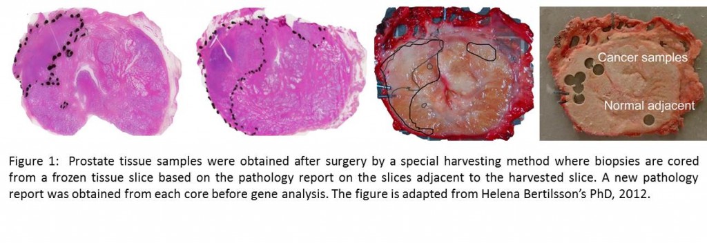
For our analysis, we used made use of a comprehensive, high quality collection of prostate cancer tissue samples stored in the Regional Biobank of Central Norway. For optimal preservation of each sample after surgery we used a standardized harvesting method previously developed in St.Olavs hospital (Figure 1). In prostate cancer, each tissue sample has its unique composition of the various tissue types: stroma, healthy epithelial cells and cancer tissue. This feature complicates our analysis by mixing the cancer-related molecular signals with signals resulting from differences in tissue composition. A strength in our material is the detailed pathology report on tissue composition attached to each sample, which makes us able to analyze the “real” cancer without the disturbance from other tissues. Such tissue correction was an important feature which helped in our discovery.
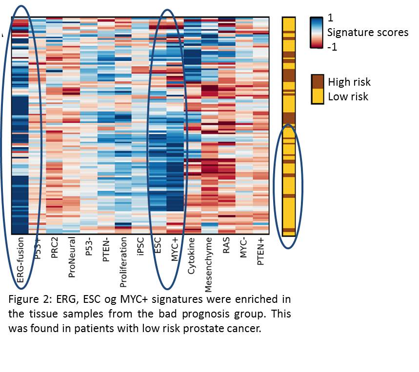
Our data highlighted two groups of samples which were related to good or bad prognosis based on survival data. The group of bad prognosis samples was characterized by enrichment of three well-known prostate cancer gene signatures (ESC, ERG and MYC+) (Figure 2). Interestingly, these signatures were found in patients diagnosed with low risk prostate cancer, addressing their “visibility” in an early stage of disease. Additionally, ESC, ERG and MYC+ were found consistently in samples from the same patient, despite highly varying tissue composition in samples from the same patient.
In our study we identified a robust molecular feature of prostate cancer related to bad prognosis, but still diagnosed as low risk cancer according to classical pathology.
Such a subtype is promising for the clinic in prioritizing these patients for radical treatment at an early stage. In future studies, we plan to validate these signatures in a larger population. We will also highlight the importance of developing methods to correct for tissue heterogeneity within each sample in molecular studies, which we found to have considerable impact on the results. Our study was recently published in BMC Medical Genomics and is currently highlighted as the editor’s first pick on the journal homepage.


