
Bloggers: Nathalie Niyonzima and Eivind Samstad
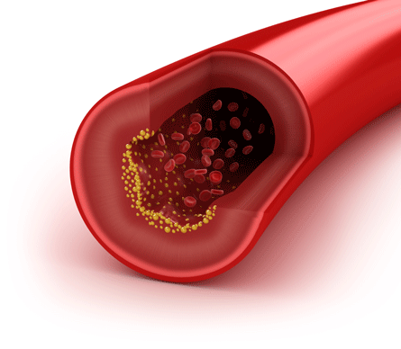
Atherosclerosis is a progressive disease that was once believed to be a disease of cholesterol storage. Today, it is well acknowledged that atherosclerosis is an inflammatory disease, and that cells and signalling molecules from the innate immune system shape the course of the disease in various ways. The defence of the normal artery depends on innate immune responses provided by endothelial cells. When challenged by inflammation, macrophages and other cells of the immune responses are recruited to the artery wall. The macrophage is an integral component to the pathogenesis of atherosclerosis, functioning at the intersection of inflammation and cholesterol homeostasis. Atherosclerotic plaque formation is driven by the persistence of lipid-laden macrophages in the artery wall. The mechanisms by which these cells become trapped, and thereby establishing chronic inflammation, remain unknown (Peter Libby, Nature 2011).
There has long been a focus on finding therapeutic methods to reduce the levels of cholesterol in the arterial wall. Studies have shown that high HDL levels are associated with reduced cardiovascular risk. This is mainly due to HDL’s ability to transport excess cholesterol in arterial macrophages to the liver for excretion (i.e., reverse cholesterol transport). Despite considerable understanding of HDL and its metabolism, therapies that aim to increase HDL levels have not been successful. Because of the heterogeneity in HDL particles, just increasing HDL levels has not been beneficial, reflecting the qualitative changes in the particles. HDL has also been shown to have other functions beyond cholesterol transport – several studies have shown that HDL is anti-inflammatory, but the mechanisms behind this are not well understood (Xuewei Zhu, Ann.Rev.Nutr 2012).
In a recent study published in the prestigious journal Nature Immunology, our research partners have taken a closer look at the anti-inflammatory effects of HDL (De Nardo, et.al, Nature 2013). They identified that HDL’s anti-inflammatory effects are mediated through the induction of ATF3. ATF3 is a key transcriptional regulator of innate immune response genes, which is induced by TLR stimulation and other stimuli, and acts as negative regulator of proinflammatory cytokines (Elisabeth S.Gold, JEM 2012).
Using mouse model and human bone marrow dendritic cells (BMDMs) treated with native HDL or reconstituted HDL prior to TLR stimuli; they showed that HDL regulates inflammation in macrophages by inhibiting transcription of proinflammatory genes such as IL-1β, IL-6 and IL-12.
In order to confirm that the anti-inflammatory effects of HDL were mediated through an inflammatory repressor, they performed microarray analysis on resting BMDMs and HDL-pretreated BMDMs subsequently stimulated with TLR ligand. ATF3 was the most induced transcription factor in the presence of HDL, and it was shown to bind to the promoters of several proinflammatory genes, thereby regulating inflammation.
They demonstrated the relevance of these findings by using Apoe-deficient mice fed a high-fat diet and injected with HDL. They observed that mice treated with HDL had lesser inflammation than the untreated ones, and that the induction of ATF3 correlated with the downregulation of proinflammatory cytokines.
Besides promoting cholesterol efflux from macrophages, HDL has been shown to protect endothelial integrity by promoting endothelial repair mechanisms. Using a model of vascular repair, they showed that the protective effects of HDL on endothelial repair are for the most part driven by ATF3.
These results provide us a link between HDL and its anti-inflammatory properties that has been a puzzle over long time. The fact that ATF3 is required for the anti-inflammatory effects of HDL shows that ATF3 is a key point for endothelial damage and inflammation. The current study has laid the foundation for understanding the regulatory mechanisms that control inflammation in atherosclerosis and other chronic inflammatory diseases.

