Blogger: Jose Teruel
PhD candidate at MR Cancer group

In a previous post in this blog I described how a new magnetic resonance imaging (MRI) technique called ‘Diffusion weighted MRI” can be applied for the detection of breast cancer due to the random movement of water molecules, or diffusion, which is different in cancer compared to healthy tissue. Therefore, using diffusion MRI we can measure the extent of diffusion and detect cancer. But, can we take advantage of this technique for benign breast lesion assessment as well?
… nowadays there is an excess of what we call ‘unnecessary biopsies’
Differentiation of breast lesions is another important step in the clinic. Let’s imagine the situation; a woman comes into the clinic feeling a lump in her breast, and a routine mammography or ultrasound confirms the presence of a suspicious lesion. The next step in the clinic would be to obtain a biopsy of the lesion to properly establish if the lesion is a malignant tumor, a threat for the patient’s life, or if it is a benign tumor that needs no further treatment.
A needle biopsy is categorized as an invasive procedure. The procedure consists in introducing a needle through the patient breast into the lesion, to obtain a sample (or several) of the tumor tissue that will be analyzed by a pathologist to examine if there is a presence of cancerous cells.
Having explained what a biopsy is, it is important to note that nowadays there is an excess of what we call ‘unnecessary biopsies’, i.e., biopsies that could have been avoided if it would have been possible to characterize the lesion as benign by a different ‘non-invasive’ procedure. Biopsies are in the best case not pleasant for the patients, and the procedure consumes time and resources.
… differentiating malignant and benign lesions without the need of an invasive procedure
So, what could the role of diffusion MRI be in this matter? Diffusion MRI can characterize different tissues based on their microstructure, and this microstructure it is known to be different in malignant and benign lesions. Our preliminary results in the use of this technique suggest a very high accuracy in differentiating malignant and benign lesions without the need of an invasive procedure. This is possible due to the fact that water movement (diffusion) is much more restricted in malignant lesions compared with benign ones and this difference can be measured by different parameters obtained from a diffusion MRI examination.
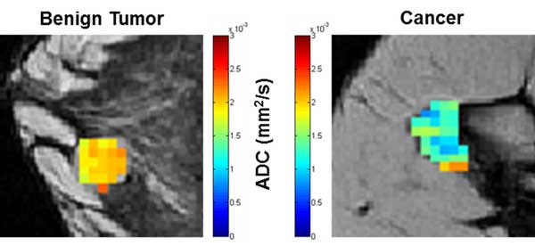
Left: Benign tumor exhibiting clearly higher values of apparent diffusion coefficient (ADC), a measure of the extent of water diffusion within tissue, compared to much lower values found for cancer on the right panel.
Furthermore, the particular technique used in our work, known as Diffusion Tensor Imaging (DTI), provides information not only in the extent of the water diffusion but also about the directionality of the diffusion. The detailed results of this work performed at NTNU and St. Olavs Hospital, is now in press and will soon be published in the Journal of Magnetic Resonance Imaging under the title “Diffusion weighted imaging for the differentiation of breast tumors: From apparent diffusion coefficient to high order diffusion tensor imaging”.
Further research and validation of our results may strengthen the current opinion that a non-invasive MRI examination can be used before biopsy to confirm if the biopsy is really necessary or if malignancy can be directly ruled out.
… diffusion MRI could save in the future a great extent of unnecessary biopsies
In summary, the use of diffusion MRI could save in the future a great extent of unnecessary biopsies avoiding invasive diagnostic procedures to patients with suspicious breast lesions.
I would finally like to thanks the dedicated volunteers that take part in our study and the funding we receive from the local health authorities and The Norwegian Research Council.