By Anna Karlberg, Department of Radiology and Nuclear Medicine, St. Olavs University Hospital, and Department of Circulation and Medical Imaging, NTNU, and Live Eikenes, Department of Circulation and Medical Imaging, NTNU.
Gliomas are the most common primary brain tumors in adults with an incidence of around 250 cases per year in Norway. The outcome is poor, and the diagnostic accuracy is essential to estimate overall prognosis and to plan the best treatment for the patient. Can a hybrid PET/MRI system improve the diagnostic accuracy and treatment planning for this patient group?
Today, the routine diagnostic work-up of glioma patients include Magnetic Resonance Imaging (MRI) and histopathological tissue sampling. Due to the heterogeneous nature of gliomas, there is however a risk of undergrading and misdiagnosis caused by sampling errors and imaging limitations.
MRI can be combined with another diagnostic technique called positron emission tomography (PET). By using radioactive tracers, PET can provide quantitative information of cellular activity and metabolism of the tumor tissue. Amino acid PET tracers are recommended as a complement to MRI by current guidelines in brain tumor imaging. This have proven promising for defining true tumor volume and differentiating viable tumor tissue from postoperative changes or radiation necrosis, and as a guide to select the best biopsy sites in gliomas. Hybrid PET/MRI therefore have the potential to improve the diagnostic accuracy compared to MRI alone.
In this study, we have used the first hybrid PET/MRI system in Norway, installed at St. Olavs University Hospital/NTNU, in the diagnostic evaluation of 11 pre-operative glioma patients. This is the first study to evaluate if the amino acid tracer 18F-fluciclovine-PET/MRI can improve the diagnostic accuracy and treatment planning in this patient group.
Fused PET/MRI and intraoperative 3D ultrasound images were used to guide surgical resection and histopathological tissue sampling in tumors with PET uptake, and image-localized histopathological samples were extracted from the tumors before resection. Analyses of the samples were related to PET/MRI image data to assess the additional value of 18F-fluciclovine-PET.
Histopathological diagnosis revealed four grade II, two grade III and five grade IV tumors. Preliminary analysis of the image data demonstrated that 18F-fluciclovine uptake was present in one grade III and all grade IV tumors, while no PET uptake was seen in grade II tumors (or grade II parts of the tumors). The tumor-to-background ratios seen in the high grade gliomas were considerably higher than that seen with other amino acid PET tracers. Preliminary results therefore demonstrate that 18F-fluciclovine PET/MRI show potential in high-grade glioma imaging particularly by guiding histopathological tissue sampling, and that 18F-fluciclovine PET might be able to differentiate between grade II and grade III tumors.
More research is however needed in this field, and future studies with amino acid-PET/MRI in patients with glioma are in the planning phase, where the main aim is to more accurately diagnose patients with this disease
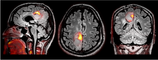
Fused MRI (greyscale image) and PET (red/yellow) images of a Grade IV glioma. The PET images demonstrated a high tumor-to-background ratio.
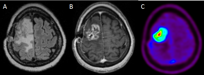
Grade IV recurrent glioma. A: FLAIR (MRI), B: Contrast-enhanced MRI, C: 18F-Fluciclovine-PET where the highest uptake of the tracer is shown in red. The PET “hot spot” can be used to take biopsies from the most malignant part of the tumor during surgery.